Conjoint Tendon Shoulder Anatomy - Laparoscopic Inguinal Hernia Repair | Basicmedical Key - Learn vocabulary, terms and more with flashcards, games and other study tools.
Conjoint Tendon Shoulder Anatomy - Laparoscopic Inguinal Hernia Repair | Basicmedical Key - Learn vocabulary, terms and more with flashcards, games and other study tools.. These are the main ligaments that help to stabilize the joints of. This video was designed for medical students depending on illustrating diagrams created by prof. What is conjoint tendon, function, definition, location and processes. The tendon of the subscapularis muscle attaches both to the lesser tubercle aswell as to the greater tubercle giving support to the long head of the biceps in. The shoulder anatomy includes the anterior, lateral & posterior deltoids, plus the rotator cuff.
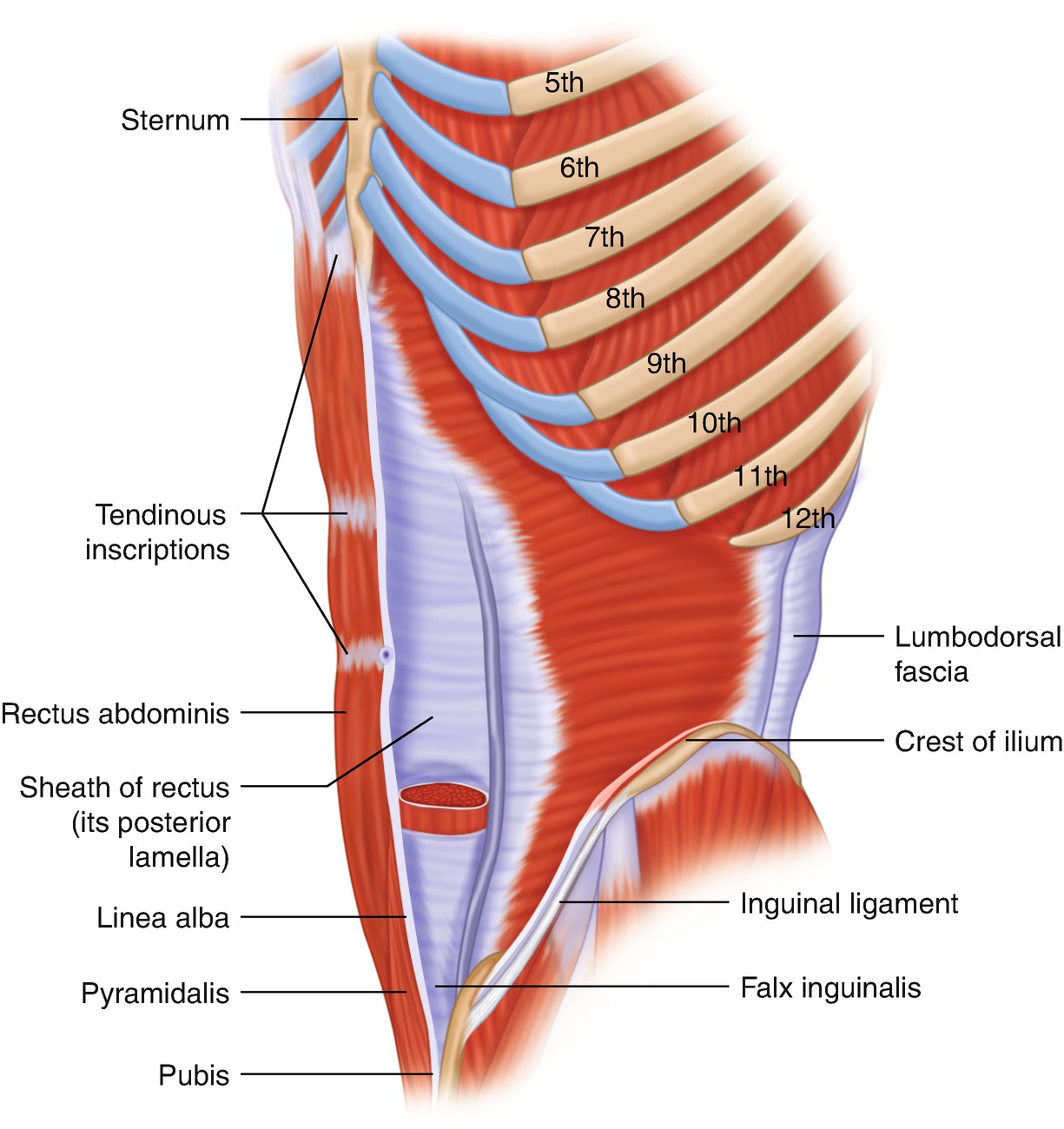
Anatomy, abdomen and pelvis, conjoint tendon (inguinal aponeurotic falx). It reduces wear and tear on the tendon during movement at the shoulder. Changes in neurovascular anatomy after open latarjet procedure 3. There are several important ligaments in the shoulder. Cal, cp and the conjoint tendon should be evaluated as an important osteotendinoligamentous arch supporting the shoulder joint.
Conjoint Tendon Shoulder Anatomy : Axilla: Anterior View ... from media.springernature.com The tendon of the subscapularis muscle attaches both to the lesser tubercle aswell as to the greater tubercle giving support to the long head of the biceps in. Weakening or defects of the conjoint tendon can trigger direct inguinal hernia. The abdominal wall is split into the posterior (back), lateral (sides). This video was designed for medical students depending on illustrating diagrams created by prof. These are the main ligaments that help to stabilize the joints of. Robin smithuis and henk jan van der woude. Qualitative and quantitative anatomy of the proximal. These are the main ligaments that help to stabilize the joints of.
Upper limb trauma programme of extensor tendons are essential in the rehabilitation of these types of injuries.
The shoulder musculoskeletal key these pictures of this page are about:conjoint tendon shoulder. Anterior projection conjoint tendon laterjet impingement. It is one of the most mobile joints in the human body, at the cost of joint stability. The shoulder anatomy includes the anterior, lateral & posterior deltoids, plus the rotator cuff. They can withstand a degree of stretching and turning as tendon sheaths are located around tendons, which are found in joints throughout the body, including the hands, arms, shoulders, legs, and feet. Normal anatomy, variants and checklist. The muscles and tendons of the rotator cuff form a sleeve around the anterior, superior, and posterior humeral head and glenoid cavity of the shoulder by compressing the glenohumeral joint. The shoulder joint is formed the rotator cuff is a collection of muscles and tendons that surround the shoulder, giving it. It reduces wear and tear on the tendon during movement at the shoulder. Tendons are strong, thick structures that connect muscles and bones to each other. There are several important ligaments in the shoulder. Anatomy, abdomen and pelvis, conjoint tendon (inguinal aponeurotic falx). Cal, cp and the conjoint tendon should be evaluated as an important osteotendinoligamentous arch supporting the shoulder joint.
Shoulder joint allows lifting, pushing and pulling by upper extremity. Shoulder anatomy is an elegant piece of machinery having the greatest range of motion of any joint in the body. Muscles allow us to move by pulling on bones. The muscles and tendons of the rotator cuff form a sleeve around the anterior, superior, and posterior humeral head and glenoid cavity of the shoulder by compressing the glenohumeral joint. Learn their origins/insertions, functions & exercises.
Scapula - Approach - Deltopectoral approach - AO Surgery ... from www2.aofoundation.org The conjoint tendon (previously known as the inguinal aponeurotic falx) is a sheath of connective tissue formed from the lower part of the common aponeurosis of the abdominal internal oblique muscle and the transversus abdominis muscle, joining the muscle to the pelvis. It is one of the most mobile joints in the human body, at the cost of joint stability. Cadaveric dissection of a right shoulder demonstrating the anatomic. Changes in neurovascular anatomy after open latarjet procedure 3. This video was designed for medical students depending on illustrating diagrams created by prof. Qualitative and quantitative anatomy of the proximal. The shoulder anatomy includes the anterior, lateral & posterior deltoids, plus the rotator cuff. Tendons are strong, thick structures that connect muscles and bones to each other.
An introduction to the anatomy of the shoulder.
Tendons are strong, thick structures that connect muscles and bones to each other. Learn their origins/insertions, functions & exercises. Anterior graphic of the shoulder. These are the main ligaments that help to stabilize the joints of. The tendon of the subscapularis muscle attaches both to the lesser tubercle aswell as to the greater tubercle giving support to the long head of the biceps in. Specifically, the four rotator cuff muscles. Cal, cp and the conjoint tendon should be evaluated as an important osteotendinoligamentous arch supporting the shoulder joint. Il rentre jeu dans la formation du… … wikipédia en français. An image depicting shoulder anatomy can be seen below. Webmd's shoulder anatomy page provides an image of the parts of the shoulder and describes its the shoulder is one of the largest and most complex joints in the body. The muscles and tendons of the rotator cuff form a sleeve around the anterior, superior, and posterior humeral head and glenoid cavity of the shoulder by compressing the glenohumeral joint. Cadaveric dissection of a right shoulder demonstrating the anatomic. Changes in neurovascular anatomy after open latarjet procedure 3.
The biceps muscle has two tendons at the shoulder, called the long head and short head. Anatomy of shoulder bones ideas shoulder anatomy medical. Magdy said to simplify the anatomical features of the. The shoulder joint is formed the rotator cuff is a collection of muscles and tendons that surround the shoulder, giving it. Coracoid process, component of conjoint tendon insertion:
Shoulder Joint Anatomy|Skeletal System|Cartilages ... from www.epainassist.com It is located in the inferior abdomen and is formed from the common aponeurosis of the internal oblique muscle and. It reduces wear and tear on the tendon during movement at the shoulder. Robin smithuis and henk jan van der woude. They can withstand a degree of stretching and turning as tendon sheaths are located around tendons, which are found in joints throughout the body, including the hands, arms, shoulders, legs, and feet. Shoulder joint allows lifting, pushing and pulling by upper extremity. These are the main ligaments that help to stabilize the joints of. Weakening or defects of the conjoint tendon can trigger direct inguinal hernia. Related online courses on physioplus.
Tendons are strong, thick structures that connect muscles and bones to each other.
The conjoint tendon, also known as the inguinal aponeurotic falx or henle's ligament, is a condensation of tissue that runs through the lateral edge of the lower rectus sheath. Anterior projection conjoint tendon laterjet impingement. The shoulder joint (glenohumeral joint) is a ball and socket joint between the scapula and the humerus. Kaddress correspondence to robert f. Changes in neurovascular anatomy after open latarjet procedure 3. Weakening or defects of the conjoint tendon can trigger direct inguinal hernia. These are the main ligaments that help to stabilize the joints of. Anterior graphic of the shoulder. In this article, we shall look at the anatomy of the shoulder joint and its important clinical correlations. Qualitative and quantitative anatomy of the proximal. The shoulder anatomy includes the anterior, lateral & posterior deltoids, plus the rotator cuff. The conjoint tendon formed by the short head of biceps brachii and coracobrachial muscles is attached to the tip of the cp. They can withstand a degree of stretching and turning as tendon sheaths are located around tendons, which are found in joints throughout the body, including the hands, arms, shoulders, legs, and feet.
Shoulder anatomy is an elegant piece of machinery having the greatest range of motion of any joint in the body shoulder tendon anatomy. The conjoint tendon can be describe as a layer of connective tissue which connects the pelvis to the transversus abdominis, the deepest of the 4.


0 Komentar